Summary
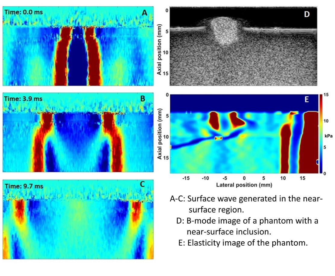
Diseases can occur inside organs and they can also occur in the near surface region of organs. Skin cancer is an example. The current shear wave elasticity imaging, as an effective method in diagnosing diseases involved in elasticity change such as cancer and liver fibrosis, is developed on bulk wave theory, in which the effect of the organ boundary on the wave propagation is not considered. One of our recent experiments shows that surface wave can be generated in the superficial region of tissue at a tissue-water interface, and it separates from the bulk shear wave during the propagation (Figure 1 A-C). Therefore, surface wave elasticity imaging method needs to be developed in order to accurately diagnose the diseases associated with elasticity variation.
Our further experiment on a tissue-mimicking phantom with an inclusion embedded in the near-surface region (Figure 1 D) show that the generated surface wave can be used for elasticity imaging (Figure 1 E). However, a comprehensive study of this method both theoretically and experimentally is yet to be performed in order to establish it as an effective and reliable method for medical diagnosis.
References
- X. M. Zhang, T. Osborn, B. R. Zhou et al., “Lung Ultrasound Surface Wave Elastography: A Pilot Clinical Study,” IEEE Transactions on Ultrasonics Ferroelectrics and Frequency Control, vol. 64, no. 9, pp. 1298-1304, Sep, 2017.