Summary
Laser-induced shear wave imaging is to utilize a pulsed laser beam to generate shear waves based on thermoelastic effects. This method can be used to measure stiffness of biological tissues noninvasively. For example, to evaluate corneal ectasia, laser irradiation was used to induce displacement at corneal surface and optical coherence tomography (OCT) was employed to detect the laser-induced displacement.1,2,3
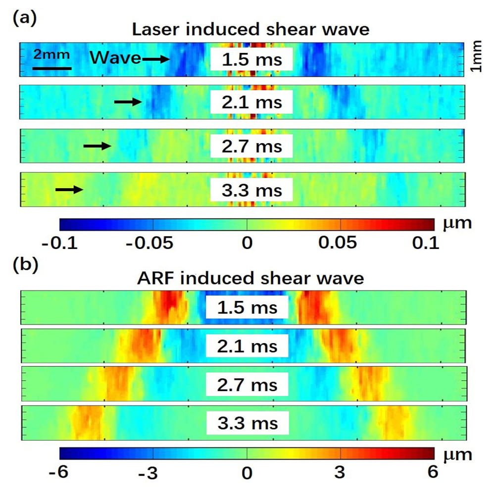
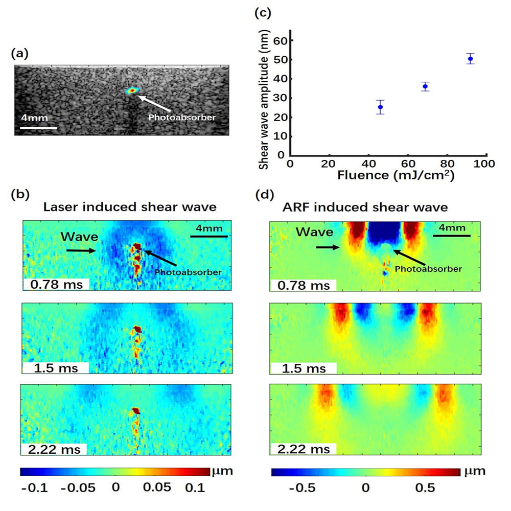
In this project, we proposed a new approach of laser-induced shear wave elasticity imaging based on optical perturbation of tissue to create shear waves and high frame rate ultrasound imaging to measure the propagation of laser-induced shear waves. In this approach, ultrafast ultrasound imaging is used to track the shear wave induced by laser irradiation. To validate the laser-induced SWEI method, we measured the shear wave velocity in the tissue mimicking phantoms and compared the results with acoustic radiation force (ARF) based SWEI. The result of this project shows that the combination of optical excitation and ultrafast ultrasound imaging of shear waves can be utilized to visualize both elastic and anatomical information of tissue with a potential in providing higher spatial resolution.
References
- C. Li, G. Guan, Z. Huang, M. Johnstone, and R. Wang, “Noncontact all-optical measurement of corneal elasticity,” Opt. Lett. 37(10), 1625-1627 (2012).^
- W. J. Dupps, “Biomechanical modeling of corneal ectasia,” J. Refract. Surg. 21(2), 186-190 (2005).^
- C. H. Liu, D. Nevozhay, H. Zhang, S. Das, A. Schill, M. Singh, S. Aglyamov, K. V. Sokolov, and K. V. Larin, “Longitudinal elastic wave imaging using nanobomb optical coherence elastography,” Opt. Lett. 44(12), 3162-3165 (2019).^