Summary
Early detection of diseases is highly depending on the spatial resolution of the imaging modality used. Although shear wave elasticity imaging (SWEI) has been proved to be an effective medical diagnostic method for many diseases including cancer and liver fibrosis and its clinical applications are continuing to expand, it is not effective in early detection of diseases. The major reason is that its current implementation, in which the same transducer is used for both shear wave excitation and tracking, make it impossible to achieve high-resolution elasticity images because of the limited bandwidth of transducers. To achieve high spatial resolution, we propose a dual-transducer approach, in which shear wave is excited using a low-frequency single-element transducer but tracked using a high-frequency array transducer.
A preliminary study was performed on a tissue-mimicking phantom of 4% gelatin containing a cylindrical inclusion with an 8% gelatin concentration. The phantom was imaged using a Verasonics Vantage ultrasound system. In the dual-transducer SWEI approach, shear waves were generated using a 5 MHz single-element transducer (Olympus, Inc.) and tracked using L22-14vLF operating at 15 MHz center frequency. These measurements were compared with those obtained using the single-transducer SWEI approach, in which shear waves were both generated and tracked using either an L7-4 at operating at 5 MHz (push/track) or an L22-14vLF operating at 15 MHz.
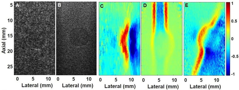
The B-scan images of the phantom with inclusion (at ~17 mm depth) obtained using L7-4 (5 MHz, Fig. 1A) and L22-14vLF (15 MHz, Fig 1B) transducers clearly show the difference in spatial resolution. The shear waves obtained using low-frequency L7-4 array transducer (Fig. 1C) were able to reach the inclusion, while high-frequency L22-14vLF array transducer could only create low amplitude shear waves in the inclusion (Fig. 1D). In contrast, our dual-transducer approach produced high amplitude shear waves at depth (Fig. 1E). The comparison of shear wave images reveals that (1) low-frequency array transducer can generate shear waves of high-intensity at deeper regions, but spatial resolution is low; (2) high-frequency array transducer can achieve high-resolution imaging but shear waves are limited to shallow depth. However, as evident from Fig. 1E, the dual transducer approach can achieve both high-resolution imaging and high penetration depth. Our results suggest that the developed approach may enable biomedical applications where high-resolution shear wave tracking is required.
References
- P. Y. Chen, C. C. Shih, W. C. Lin et al., “High-Resolution Shear Wave Imaging of the Human Cornea Using a Dual-Element Transducer,” Sensors, vol. 18, no. 12, Dec, 2018.